A Clinicians Reference Guide to Marburg Virus Disease

Share this article
Reports of Marburg virus disease (MVD) outbreaks in Equatorial Guinea on February 13, 2023 (WHO, accessed 2/24/23, https://www.afro.who.int/countries/equatorial-guinea/news/equatorial-guinea-confirms-first-ever-marburg-virus-disease-outbreak), and in Ghana last August are just the latest in the 50-year history of this deadly viral hemorrhagic fever. Beginning with the first reports of infection in laboratory workers in 1967, MVD has re-emerged repeatedly. Yet there are no medical countermeasures (e.g., vaccines or therapeutics) licensed to fight MVD, which has a case fatality of up to 88 percent.
Global health care and public health systems need to boost preparedness: improving surveillance, expanding diagnostic capacity, implementing infection prevention and control measures, as well as intensifying efforts to identify effective medical countermeasures.
Published in 2020, “Marburg virus disease: A summary for clinicians,” summarizes MVD countermeasures, focusing on pathogenesis, clinical features, and diagnostics, with an emphasis on therapies and vaccines that have demonstrated potential for use in an emergency situation, through their evaluation in non-human primates (NHPs) and/or in humans.
Here, we provide a summary of key features of MVD from the manuscript, and updates on therapeutics and vaccines in development.
10 Takeaways for Clinicians for Marburg Virus Disease (MVD)
- MVD is a rare but severe viral hemorrhagic fever caused by Marburg virus (MARV), which is a filovirus (CDC, https://www.cdc.gov/vhf/virus-families/filoviridae.html, Accessed 2/24/2023) and closely related to Ebola virus.
- Cases of MVD have primarily occurred in sub-Saharan Africa, and the transmission risks, clinical features, and high case fatality rates are similar to Ebola virus outbreaks.
- Marburg virus infection can occur after exposure to the droppings or urine of African fruit bats or bodily fluids from infected animals. Person-to-person transmission occurs in households and health care settings via contact with the infected individual’s blood or bodily fluids, or from touching contaminated surfaces and materials. Learn more about personal protective equipment (PPE) recommendations when caring for a person with suspected or confirmed MVD.
- MVD has an incubation period of about 3-21 days, after which sudden symptom onset usually occurs. Early symptoms of MVD may look like more common febrile illnesses such as malaria and typhoid, which can result in missed or delayed diagnosis of MVD. There have been no documented asymptomatic cases of MVD.
- A detailed exposure history, including travel to endemic areas, contact with sick persons or animals, activities during the previous 3 weeks including visiting caves, and other risk factors should be obtained to identify patients with recent travel and compatible symptoms who may be at risk for MVD.
- Health care personnel (HCP) who identify a patient with suspected or confirmed MVD should follow an identify, isolate, and inform strategy and implement appropriate isolation and personal protective equipment. See NETEC’s Identify, Isolate, and Inform Tip Sheet.
- If MVD is suspected, diagnostic testing must be coordinated with local public health officials and the Centers for Disease Control and Prevention (CDC). Contact the CDC (CDC, https://www.cdc.gov/ncezid/dhcpp/vspb/specimens.html, Accessed 2/24/2023).
- Early clinical presentation of MVD may include fever, chills, malaise, headache, muscle aches, vomiting, and diarrhea. Maculopapular rash is an early sign of MVD. Progression to severe MVD may lead to decreased alertness, bleeding and bruising, hypotension, shock, and multiorgan failure.
- Management of patients with MVD is primarily supportive care in the hospital setting. Rehydration with oral or intravenous fluids and treatment of specific symptoms improves patient outcomes.
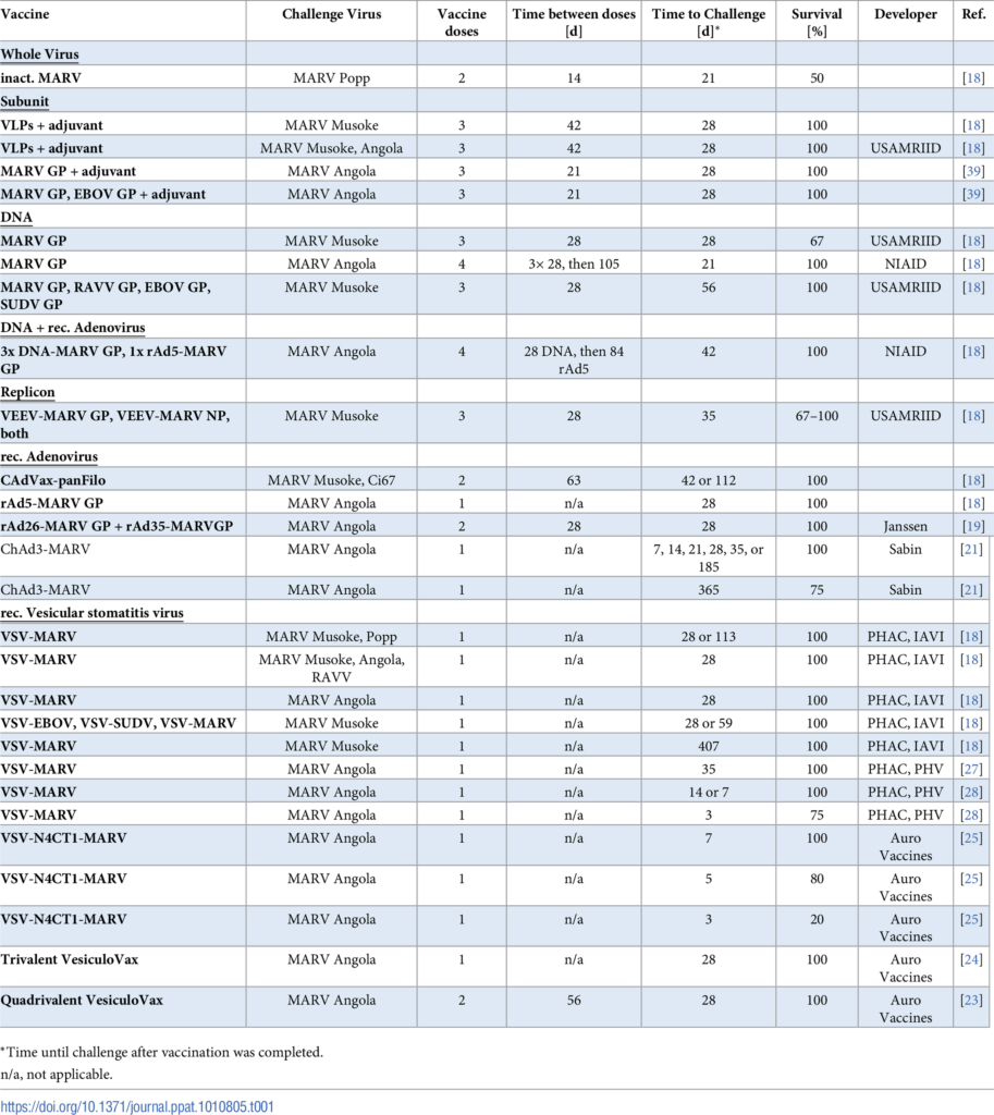
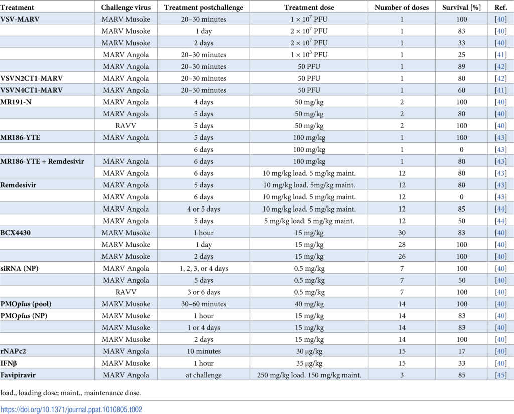
- Currently there are no approved medical countermeasures for MVD, but several vaccine candidates for use as pre- and post-prophylaxis are in preclinical development and two have undergone Phase I clinical testing. Treatment options for MVD have not been definitively established, but multiple therapeutics such as monoclonal antibodies (mAb) and antivirals have been tested in non-human primate models of disease. The World Health Organization recently hosted a review of current state of MVD therapeutics (WHO, accessed 2/24/23, https://cdn.who.int/media/docs/default-source/blue-print/simon-funell_whomarvac_therapeutics_14feb23.pdf?sfvrsn=bcf02d72_3) including mAb therapies, post-exposure prophylaxis, and antivirals and vaccine candidates (WHO, accessed 2/24/26, https://cdn.who.int/media/docs/default-source/blue-print/william-dowling_whomarvac_vaccines_14feb23.pdf?sfvrsn=df284d46_3).
MVD Therapeutics and Vaccine Candidates
The WHO R&D Blueprint has convened a group of experts in the field of vaccine research and development to promote development of Marburg virus (MARV) vaccine candidates. The following tables were produced by the MARV vaccine “MARVAC” consortium (https://journals.plos.org/plospathogens/article?id=10.1371/journal.ppat.1010805, accessed 2/24/23).
Additional Resources
About Marburg Virus Disease (CDC, https://www.cdc.gov/marburg/about/, accessed 2/24/23)
Marburg Virus Disease Fact Sheet (WHO, https://www.who.int/news-room/fact-sheets/detail/marburg-virus-disease, accessed 2/24/23)
About the Author
Athena Plichta, BSN, RN, DTN, a-IPC, is a public health nurse and graduate student in the MPH-Epidemiology program at the University of Nebraska Medical Center (UNMC). She is interested in emerging infectious diseases, infectious disease epidemiology and preparedness, infection prevention and control, and biodefense. She currently works at a local health department in Northern Virginia and is a student worker at NETEC.
This post was reviewed by NETEC subject matter experts Erica Shenoy, MD, PhD, and Corri Levine, PhD, MS, MPH.